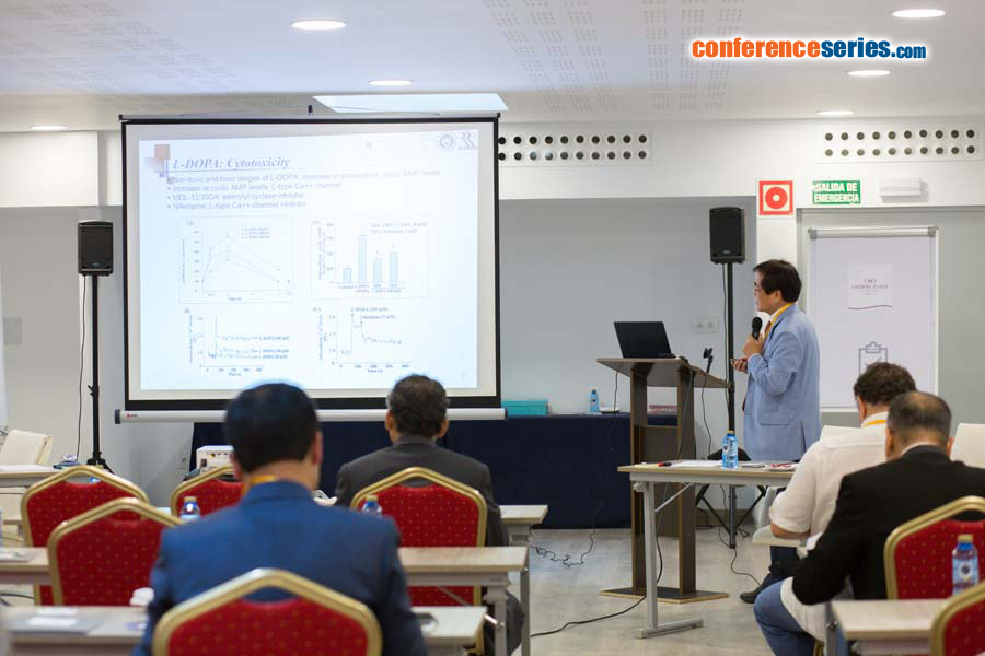
Myung Koo Lee
Chungbuk National University, Republic of Korea
Title: Modulation of cell viability by L-DOPA via the ERK and JNK-c-Jun systems in dopaminergic neuronal cells
Biography
Biography: Myung Koo Lee
Abstract
Parkinson’s disease (PD) is caused by the degeneration of dopaminergic neurons in the substantia nigra-striatal region. L-3,4-dihydroxyphenylalanine (L-DOPA) is the most frequently prescribed drug for controlling the symptoms of PD. However, the high levels of L-DOPA lead to cell death by generating reactive oxygen species in dopaminergic neuronal and PC12 cells. Recently, it has been reported that the intracellular cyclic AMP levels increased in response to cytotoxic levels of L-DOPA and multiple treatments with non-toxic L-DOPA (MT-LD) reduced dopamine biosynthesis in PC12 cells. In this study, the effects of L-DOPA on dopaminergic neuronal cell death via the ERK1/2 and JNK1/2-c-Jun systems were investigated. In PC12 cells, MT-LD (20 μM) induced cell survival via PKA-transient ERK1/2 activation, and then it induced differentiation via the Epac-sustained ERK1/2 system. MT-LD initially enhanced c-Jun phosphorylation (Ser73), but later induced c-Jun phosphorylation (Ser63) and c-Jun expression, which subsequently led to the cell death process. In the 6-hydroxydopaminelesioned rat model of PD (6-OHDA lesion), L-DOPA administration (10 mg/kg) protected against neurotoxicity through c-Jun phosphorylation (Ser73) for 1–2 weeks. However, L-DOPA administration (10 or 30 mg/kg) showed neurotoxicity through c-Jun phosphorylation (Ser63) and c-Jun expression via ERK1/2 phosphorylation for 3–4 weeks. In addition, gynosaponin TN-2 from ethanol extract of G. pentaphyllum (GP-EX) protected against L-DOPA-induced neurotoxicity in PC12 cells. Gypenosides and GP-EX also showed the protective effects on long-term L-DOPA administration in 6-OHDA lesion. Our data indicate that chronic treatment of L-DOPA causes neurotoxicity via the cyclic AMP-ERK1/2-c-Jun system in dopaminergic neuronal cells and GP-EX might serve as an adjuvant agent for PD.
Recent Publications :
1. Shin K S, Park H J, Park K H, Lee K S, Jeong S W, Hwang B Y, Lee C K and Lee M K (2018) Eff ects of gynosaponin TN-2 on L-DOPA-induced cytotoxicity in PC12 cells. Neuro Report 29(1):1-5.
2. Zhao T T, kim K S, Shin K S, Park H J, Kim H J, Lee K E and Lee MK (2017) Gypenosides ameliorate memory defi cits in MPTP-lesioned mouse model of Parkinson's disease treated with L-DOPA. BMC Complementary and Alternative Medicine 17(1):449.
3. Park K H, Shin K S, Zhao T T, Park H J, Lee K E and Lee M K (2016) L-DOPA modulates cell viability through the ERKc- Jun system in PC12 and dopaminergic neuronal cells. Neuropharmacology 101:87-97.
4. Shin K S, Zhao T T, Park K H, Park H J, Hwang B Y, Lee C K and Lee M K (2015) Gypenosides attenuate the development of L-DOPA-induced dyskinesia in 6-hydroxydopamine-lesioned rat model of Parkinson's disease. BMC Neuroscience 16:23.
5. Shin K S, Zhao T T, Choi H S, Hwang B Y, Lee C K and Lee M K (2014) Eff ects of gypenosides on anxiety disorders in MPTP-lesioned mouse model of Parkinson's disease. Brain Res 1567:57-65.


